Discoveries
MESA Researchers Learn how to Predict Risk of Heart Disease Better...And win an Award!
An article based on data collected in the Multi-Ethnic Study of Atherosclerosis
(MESA) was recently recognized as the best original scientific article published
in the journal Radiology! The annual award, called the “Alexander R. Margulis Award for Scientific Excellence,”
is given to the article which best exemplifies high levels of scientific quality and originality.
MESA researchers, led by Drs. Anna E.H. Zavodni and David A. Bluemke, earned the award this year because they asked the question:
is there a better way we can predict who is at risk of heart disease?
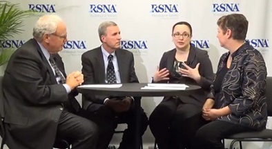
The authors discuss their research with the editors of the journal Radiology.
From left to right: Herbert Y. Kressel, MD (Editor of Radiology);
David A. Bluemke, MD, PhD; Anna E. H. Zavodni, MD; Deborah Levine, MD (Deputy Editor of Radiology).
(Image Copyright ©2010 Radiological Society of North America, Inc. (RSNA), All Rights Reserved)
Scientists and doctors already know about some things that make a person more likely to develop heart disease,
like high blood pressure and cholesterol. MESA Investigators are trying to learn more about atherosclerosis.
Atherosclerosis, commonly thought of as the “hardening of the arteries,”
is the end result of cholesterol deposition in the artery wall, forming “plaque” thickening of the artery wall.
If the plaque ruptures into the blood stream, it might cause a stroke or a heart attack.
Unfortunately, many times there’s no obvious indication that a person is developing atherosclerosis until it causes
a problem—like a heart attack. That’s why MESA researchers are working hard to see if there are ways to predict
disease before it becomes a problem, so attempts at prevention can be made. (Would you like to learn more about atherosclerosis?
Check out this page for a step-by-step animation: http://watchlearnlive.heart.org/CVML_Player.php?moduleSelect=athero).
|
Different features of the plaque can tell us how dangerous it is (in other words, how likely it is to rupture).
We can look at these different features of the plaque using magnetic resonance imaging, or MRIs.
MRIs use powerful magnets to take pictures of tissues inside the body, and they are painless and noninvasive.
|
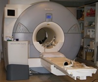
An MRI machine
|
MESA collaborators wondered if they could use MRI pictures of arteries to look at plaques and see if they
could predict who might be at risk for heart disease in the future.
Dr. Bluemke and Dr. Lima at the MESA MRI reading center have been working on this question through the MESA study
for over a decade. Dr. Zavodni took on the project early in her career, under the supervision of Dr. Bluemke.
The researchers took pictures of the carotid arteries, which are blood vessels in the neck that help bring oxygen
to the head (see picture below). Scientists think the carotid artery is a pretty good representation of what the rest
of the blood vessels in the body look like. The carotid artery is also helpful because it is pretty large compared
to the other arteries and it is also fairly close to the surface of the skin. This makes it easier for researchers to
capture the images they need.
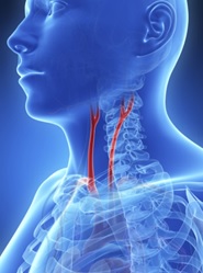
Carotid arteries
MESA researchers were able to look at MRI pictures of the carotid arteries of 946 participants.
Then they kept in touch with those participants for about five years. After five years, 59 (or 6%)
of those 946 participants had experienced some sort of cardiovascular event, like a heart attack or a stroke.
The scientists compared the pictures of the arteries of the people who did have an event to the pictures of the people who didn’t have an event.
Were they different? If so, the pictures might be able to help us predict who is at risk of events like heart attacks. And yes!
The pictures were different. The scientists were able to see things on the pictures, like how thick the walls of the arteries were,
that seem to be linked to that person’s risk of events like heart attack or stroke.
Check out the picture of the carotid artery at the bottom of the page for a visual example of the impact of atherosclerosis on arteries.
This is great news because this information can be used by doctors to predict a patient’s risk level more accurately than
with traditional risk factors, like high blood pressure, high cholesterol, obesity, and cigarette smoking, etc.
In fact, of the 59 participants in the study who experienced an event, 16% more of them could be identified as at risk with
the artery imaging technique than could be identified with just the traditional risk factors. With the earlier recognition
of risk that the artery images can provide, we can hope to help prevent heart disease by implementing lifestyle changes or treatment.
Congratulations to Drs. Zavodni and Bluemke and their team for this well-deserved recognition,
and thank you to all of our participants, whose dedication helped make this discovery possible.
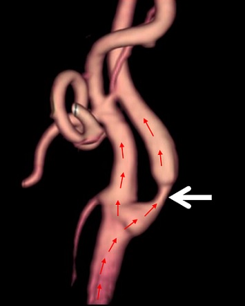
MRI of the carotid artery.
Blood flows from the heart up the artery (direction and path of blood flow is indicated by the small red arrows),
supplying the brain, facial region, and eyes.
At the region of the large white arrow, the artery is severely narrowed due to atherosclerotic
plaque in the surrounding artery wall.
References:
http://www2.rsna.org/timssnet/media/pressreleases/14_pr_target.cfm?ID=802
http://pubs.rsna.org/page/radiology/margulis
http://www.ncbi.nlm.nih.gov/pubmed/24592924